185 Alewife Brook Parkway, suite 210
Cambridge, MA 02138
USA
CYTOO TECHNOLOGY
CYTOO Technology is a perfect compromise between conventional conditions that introduce variability in cell functions, and multilayer conditions that implies very complex image analysis.
In vivo, cell behavior results from the integration of different signals obtained from adhesion molecules, paracrine and endocrine mediators and from mechano-structural guidance, a relationship that has been largely ignored in conventional culture systems. Conventional in vitro cellular assays are performed on uniform adhesive substrates that can introduce considerable but unnoticed variability in cell function.
CYTOO Technology provides structural guidance to cells cultured in vitro and thus the framework for expression of a more physiological phenotype. Guidance can improve experimental reproducibility, sensitivity and provide a tool for understanding the relationship between structure and function in quantifying complex biological events. CYTOO restores the possibility for cells to adapt to their boundary conditions thus returning the opportunity to behave as mechano-sensing systems.
Structural guidance is brought by adhesive micropatterns, where size, shape and nature of the adhesive surface can be fine-tuned to best fit the needs of your assay.
BACKGROUND
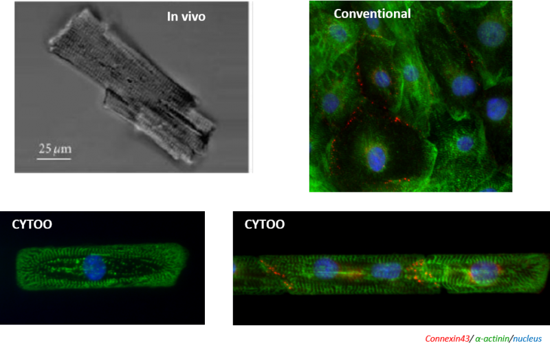
Micropatterns allow you to reconstitute in vitro some in vivo-like conditions. For example, cardiomyocytes isolated from a healthy human heart present a brick-like morphology with nicely aligned sarcomeres (Harvey and Leinman, 2011). By controlling cell adhesion using micropatterns, one can restore some of the physical constraint present in vivo, not only at the single cell level but also by organizing multiple cells. In this example, one can observe that micropatterned cardiomyocytes have both cell and sarcomeres aligned, reconstituting the in vivo situation. Such morphology is completely lost when cardiomyocytes are plated on conventional petri dishes. In addition, cardiomyocytes on micropatterns show increased levels of connexin-43 compared to standard culture conditions, showing that the restored microenvironment truly impacts on gene expression and the models magnitude of maturity.
MICROPATTERNS
CYTOO’s micropatterns are produced by a process called photolithography in which a non toxic cell repellent surface (called cytophobic surface) is degraded locally by the UV light passing through a photomask.
Micropatterns are then coated with dedicated ECM protein to form adhesive substrates of defined areas dedicated to cell-culture, surrounded by a cytophobic surface. They allow a controlled and standardized attachment and spreading of cultured mammalian cells. Micropatterns reproduce micro-environmental cues that control the shape, organization, polarity, spreading and internal organization of attached cells, through a strong impact on genome transcription.
MORE PHYSIOLOGICALLY RELEVANT CELLULAR MODELS
Over the last decade, work performed by many research groups has shown how the tight control of adhesive and non-adhesive surfaces on micropatterns can normalize cell organization and behavior. Like in tissues, cells have a highly reproducible response when constrained by such boundary conditions.
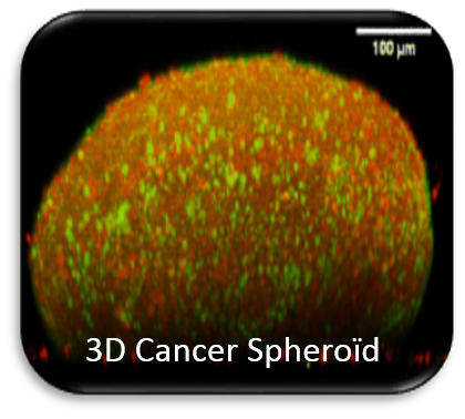
Using its acquired expertise on controlling cell behavior of single cells, CYTOO has dedicated its R&D to the development of innovative cellular models and readouts with the constant objective of higher physiological relevance. These developments include multicellular models for key applications in toxicology as well as spheroids for oncology. In each of these fields, combining physiological relevant cells with a controlled micro environment translates into higher maturation or differentiation while maintaining compatibility with automation and High Content Screening and Analysis.